Space Ranger2.0, printed on 03/11/2025
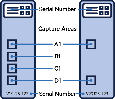
The microscope slides used to generate Visium data have features that Space Ranger users need to know. In order to run Space Ranger, it is necessary to know the serial number associated with the slide that generated the data you intend to analyze, as well as the capture area associated with each sample.
The image below illustrates these key features of the Visium slide and these terms are explained in the glossary below.
 |
 |
 |
 |
Alignment File - A file produced by the Loupe Browser when using either manual fiducial alignment with tissue detection or manual image registration.
Area (or Capture Area) - One of the either four or two active regions where tissue can be placed on a Visium slide. Each area is intended to contain only one tissue sample. Slide areas are named consecutively from top to bottom: A1, B1, C1, D1 for Visium slides with 6.5 mm Capture Area and A, B for CytAssist slides with 11 mm Capture Area. Both CytAssist slides with 6.5 mm Capture Area and Gateway Slides contain only two slide areas, A1 and D1. More information on the slide parameters page.
Brightfield Image - A light-microscopy image depicting tissue on a slide. In a Visium experiment, a brightfield image is used as an anatomical reference. These images are usually stained with hematoxylin and eosin in order to highlight tissue structure (see H&E staining below).
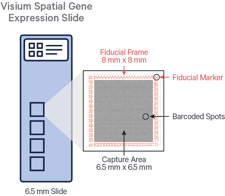
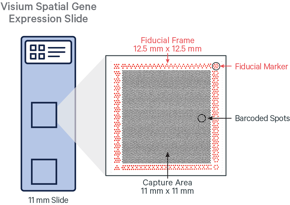
Barcoded Spots - The invisible spots on the slide that contain specialized oligos for capturing poly-adenylated mRNA for Gene Expression .
Fluorescence Imaging - An imaging technique whereby signal is generated by fluorophores that re-emit narrow-spectrum light when excited by light of a certain wavelength or color.
Immunohistochemistry (IHC) and Immunofluorescence (IF) - IHC is an imaging technique whereby protein is stained using a matching, antibody-based label. Immunofluorescence (IF) is a subset of this technique that generates a signal using fluorophores attached to antibodies and fluorescence imaging.
Fiducial Frame - A frame of specially patterned spots surrounding each capture area. These spots help the sample microscopist see where to place tissue and are also used by Space Ranger to determine where the capture area is in an image.
Fiducial Marker - A subset of the fiducial spots in each corner of the capture area that make easily identifiable shapes including an hourglass, a triangle, an open hexagon, and a filled hexagon.
Gateway Slide - A Visium Spatial Gene Expression slide with a reduced number of capture areas. These slides are the same as the original Visium slides, but only have capture areas A1 and D1. Gateway slides have serial numbers starting with "V2".
H&E Staining - The process of applying hematoxylin and eosin to tissue in order to highlight tissue structure. Hematoxylin colors cell nuclei blue, and eosin colors the cytoplasm and extracellular matrix pink.
Sample - A single tissue section applied to a single capture area on a Visium slide.
Slide File - A file describing the layout of capture spots in a single slide identified by slide serial number.
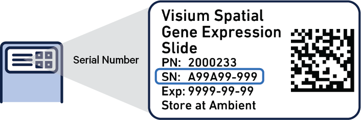
Slide Serial Number - The unique identifier printed on the label of each Visium slide. The serial number starts with V followed by a number which can range between one through five and ends with a dash and a three digit number, such as 123.
Count Matrix (or Feature-Barcode Matrix) - A matrix of counts representing the number of unique observations of each feature (gene) within each Visium spot barcode. Genes defined by the transcriptome reference and antibodies defined in the Feature Reference appear as rows in the matrix. Each barcode is a column of the matrix.
GEX: Short hand notation for Gene Expression, which is a process by which the information encoded in a gene is transcribed to either RNA molecules that code for proteins.
Dual Indexing - A strategy for sequencing multiple samples on the same flowcell by using two oligonucleotide sequences, one attached to either end of each fragment to be sequenced, in order to uniquely identify the sample. The Visium library construction only supports multiplexing samples using this dual-index strategy. See sample index below.
Library (or Sequencing Library) - A Visium spatially-barcoded sequencing library prepared from a single slide area.
Sample Index - An oligonucleotide sequence used in library construction to differentiate multiple samples that are sequenced on the same flowcell. On the Illumina platform, these sequences are read out as separate "index reads" and reads are sorted into sample-specific files using mkfastq. The Visium library construction supports only "dual-indexing" (see above).
Sequencing Run (or Flowcell) - The output data, including Illumina BCL files, from one sequencing instrument run. The data can be demultiplexed by lane or by sample index. See mkfastq for more information about demultiplexing.
Target Panel CSV file - A CSV file declaring the target gene panel used for a Targeted Gene Expression experiment, which specifies detailed information about the targeted genes and baits included in the panel. This file must be provided to spaceranger count via the --target-panel option when analyzing Targeted Gene Expression data. Details and specifications of the Target Panel CSV file format are documented here.
Baits - A set of oligonucleotide sequences designed to specifically hybridize to and recover molecules from targeted genes during the hybridization-capture enrichment step of the Targeted Gene Expression assay.
WTA (or Whole Transcriptome Analysis) - Whole Transcriptome Analysis, describes a (non-targeted) Visium spatial RNA-seq library or dataset which may be used as input for a Targeted Gene Expression experiment.
Parent - A WTA spatial gene expression library or dataset that was used as the input material for a Targeted Gene Expression experiment. Because the Parent and its corresponding Targeted dataset are both derived from the same Library, it is possible to directly compare results from these matched datasets in a pairwise manner, as enabled by spaceranger targeted-compare.
Probe Barcode: The unique barcode on the right hand side (RHS) probe.
Probe Set - A whole-transcriptome reference file declaring the gene panel used for a Visium for FFPE experiment, which specifies detailed information about the genes which are targeted by each probe. This file must be provided to spaceranger count via the --probe-set option when analyzing Visium for FFPE data.
Probe Filter - Within the whole-transcriptome Probe Set reference file declaring the gene panel used for a Visium for FFPE experiment there is an included column. By default, probes predicted to have some off-target activity to homologous genes or sequences are excluded from analysis. Users can include UMI counts from all probes, including those with potential off-target activity, with use of the --filter-probes=false option which is true by default.
RNA-Templated Ligation - Visium for FFPE is designed around a strict probe pairing framework. For each target locus, when two half-probe sequences bind to the proper locus and ligate together during the assay, a countable barcode-UMI-probe product is made.
Visium CytAssist Instrument: An instrument that mediates the tissue permeabilization to release the ligation products from tissues on standard glass slide for capture by spatially barcoded oligonucleotides within each Capture Area on the Visium slide. It also captures the image of the tissue section on the Visium slide.
CytAssist Captured Image (or CytAssist Image): A low resolution brightfield image in TIFF format that is captured by the CytAssist of the eosin stained tissue section on the CytAssist slide inside the instrument. This image contains the fiducial frame.
Microscope Image: A high resolution brightfield or fluorescence image of the tissue section on the standard glass slide captured by a microscope. This image does not contain the fiducial frame.
CytAssist Spatial Gene Expression Slide, 6.5 mm: Visium Spatial Gene Expression slide for use with CytAssist instrument with two capture areas each with dimensions of 6.5 mm by 6.5 mm. The spots within the capture area on these slides contain specialized oligos for capturing poly-adenylated mRNA tags. These slides have serial numbers starting with "V4".
CytAssist Spatial Gene Expression Slide, 11 mm: Visium Spatial Gene Expression slide for use with CytAssist instrument with two capture areas each with dimensions 11 mm by 11 mm. The number of spots within the capture area on these slides are ~3x higher compared to the 6.5 mm capture areas and contain specialized oligos for capturing poly-adenylated mRNA tags. These slides have serial numbers starting with "V5".